Scientists create first nanoparticle-based tissue adhesive
July 21, 2017 | Friday | News
Its properties were successfully tested in sealing a liver puncture and in conducting operations in moving organs, like lung and limbs.
Courtesy- Wikimedia
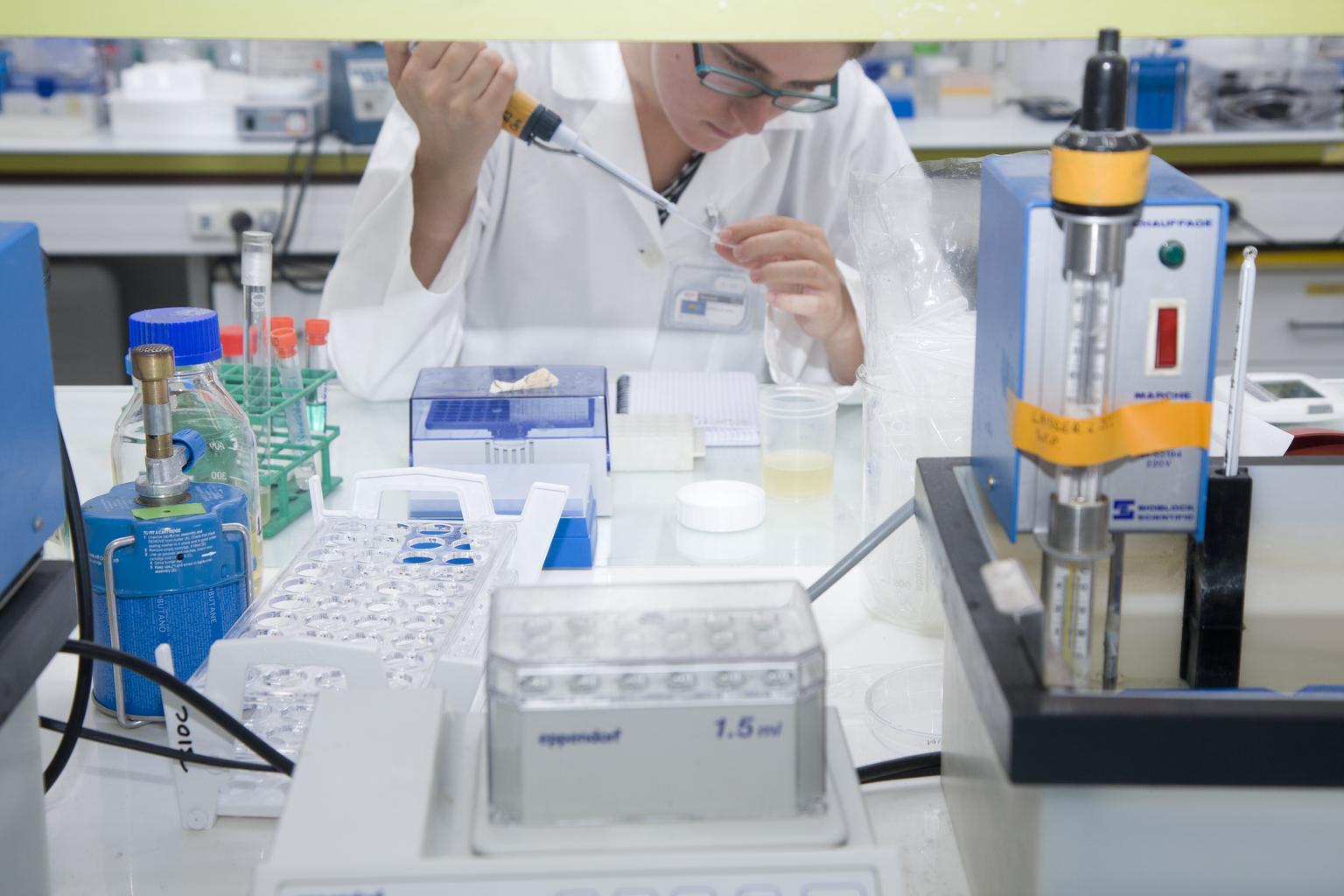
Researchers at the Center for Nanoparticle Research, within the Institute for Basic Science (IBS) in collaboration with medical doctors in Seoul National University Hospital, created a surgical glue that is both adherent and visible in the most common imaging techniques: fluoroscopy, ultrasound, and computed tomography (CT).
It is the first nanoparticle-based tissue adhesive that features these characteristics. Its properties were successfully tested in sealing a liver puncture and in conducting operations in moving organs, like lung and limbs.
IBS scientists in collaboration with researchers at Kookmin University and Seoul National University Hospital designed nanoparticles with a shell made of silica (SiO2) and a core of radiopaque tantalum oxide (TaOx). SiO2 holds the tissue together, while TaOx provides contrast enhancement on ultrasound and CT.
The research team tested the TaOx/SiO2 core/shell nanoparticle (TSN) glue and found that it is clearly visualized by real-time imaging modalities and exhibits adhesive properties similar to that of the U.S. Food and Drug Administration (FDA)-approved cyanoacrylate and Lipiodol (CA-Lp), a mixture of a tissue adhesive and radiopaque oil used in the clinical practice.
This is important because it could help surgeons to recognize a moving surgical target and perform a safe and accurate operation.